Jan 20,2021|By
Recently, a research group led by Prof. WANG Junfeng from the High Magnetic Field Laboratory, Hefei Institutes of Physical Science, made a progress in the research of biological protein template regulating the growth of nanocrystals and performed some applications.
Magnetic resonance contrast agent can improve the image resolution of magnetic resonance imaging (MRI) by changing the signal intensity of biological tissue through internal and external relaxation effect and magnetic susceptibility.
MRI contrast agent can be divided into positive (T1) and negative (T2) contrast agent according to the enhancement type. Gadolinium based contrast agent (Gd-DTPA), which is commonly used in clinic, may release a small amount of free Gd ions in vivo, resulting in serious toxicities and side effects.
Iron oxide nanoparticles with ultra-small size (less than 5 nm) have good biocompatibility and are considered to be an ideal substitute for gadolinium based contrast agents.
However, the current preparation of small-size iron oxide nanoparticles is usually synthesized in organic solvents, and further hydrophilic ligand replacement is therefore needed, which is expensive and environmentally harmful, resulting in increased difficulty and reduced efficiency of synthesis. Exploring iron oxide nanoparticles with good biocompatibility and high T1 contrast effect in a facile and efficient method remains a great challenge.
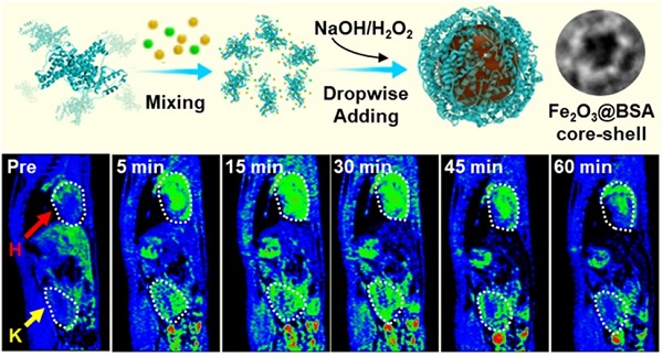
In this research, the ultra-small size Fe2O3@BSA nanoparticles (3.5 nm) were successfully synthesized with highly uniformity and monodispersity, which was regulated by the cage-like protein structure self-assembled of bovine serum albumin (BSA).
Compared with the commercially available Gd-DTPA (Magnevist), Fe2O3@BSA nanoparticles have brighter signal and longer angiographic effect, indicating a great potential as a T1 contrast agent in steady-state and high-resolution MR imaging.
In addition, researchers found that, owing to the BSA coating, the nanoparticles can be metabolized in vivo and excreted through urine, which has good biological safety and great commercial prospects.
The results, entitled “In Situ One-Pot Synthesis of Fe2O3@BSA Core-Shell Nanoparticles as Enhanced T1-Weighted Magnetic Resonance Imagine Contrast Agents”, were published in the ACS Applied Materials & Interfaces.
This research was supported by grants from the Ministry of Science and Technology of China and the National Natural Science Foundation of China.

The BSA cage-like nanoreactor and MR images in vivo
Attachments Download: